Evaluating the Efficacy of the 3D U-Net Architecture For Glioblastoma Multiforme Tumor Segmentation
This research evaluates the performance of the 3D U-Net model for automated glioblastoma tumor segmentation from MRIs, achieving 98.6% accuracy and significantly faster processing times than human oncologists, crucial for effective radiation therapy.
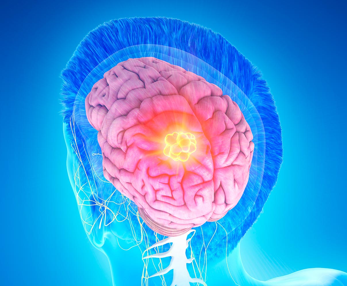
Glioblastoma is the deadliest form of brain cancer which begins as a congregation of cancerous cells within the brain but then progresses into invading and destroying healthy brain tissue [9]. Radiation therapy, the most popular treatment option, is where neuro-oncologists apply intense radiation energy beams directly on the tumor region to kill the cancerous cells. However, for radiation therapy to be effective, the segmented magnetic resonance image (MRI) in which the oncologists base where to apply the radiation must be segmented nearly 100% accurately, or else the energy beams will mistakenly damage healthy brain tissue. Additionally, radiation therapy will be futile if the MRI segmentation is not complete in time for the therapy appointment. Our research problem is evaluating the performance of notable segmentation models for accurate and immediate 3D segmentation of glioblastoma multiforme brain tumors from MRIs while focusing on one particular architecture, the 3D U-Net. Each of the models was given 850 MRIs from the BraTS 2021 dataset, which is an annual competition hosted by the University of Pennsylvania [8]. Our 3D U-Net model achieved a testing accuracy of 0.986) accuracy and took approximately 55 seconds to predict segmentations of the tumor region. The results reveal that the 3D U-Net model is capable of automating glioblastoma tumor segmentation in significantly fewer hours than a human oncologist would take, all while maintaining similar or higher accuracy, where even a minor difference can mean the distinction between life and death.
Explore More!

Related Projects

© 2022 Inspirit AI. All Rights Reserved.