Back To Projects

Detecting Retinal Detachment using images with Machine Learning

Yi-Chen (.

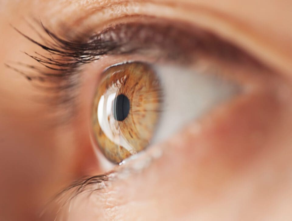
This research is on how AI and machine learning can be used to detect retinal detachment using images.


Retinal detachment occurs when your retina (a light-sensitive layer of tissue in the back of your eye) is pulled away from its normal position at the back of your eye. Symptoms include increased eye floaters, sudden flashes of light in vision, and dark shadows in the middle or on the sides of vision. It can eventually lead to blindness. Therefore, it is important that we detect it early on and treat it. This research is on how AI and machine learning can be used to detect retinal detachment using images. Before building my models, I did data pre-processing on retinal images data from four different hospitals. Then I built several different machine learning models, including Logistic Regression, Ridge Classifier, Decision Tree Classifier, K-nearest neighbor, Random Forest Classifier, SVM, MLP Classifier, Neural Networks, and Transfer Learning. I then assessed their accuracy using certain metrics. After building the models, I calculated the accuracy score, precision score, recall score, F1 score, and built Confusion Matrices for all of the models. Our results showed a very high accuracy in all of them; they were all in the 90% to 99% range. Therefore, I can assume that Retinal Detachment can be diagnosed using Machine Learning methods. In summary, retinal detachment is a serious issue and needs to be detected and treated. Using our machine learning models, I achieved great results.

Explore More!

Yi-Chen (.
Samar Abu Hegly
BS in Electrical Engineering & Computer Science and Neuroscience from MiT

Related Projects

workspace_premium
Differences in predicted rates of vaginal births after cesarean across racial groups in a ‘race-neutral’ model

A large body of work in machine learning has highlighted that supposedly de-biased systems often re-code sensitive variables like race in terms of proxy variables. In order to determine if this was the case in this calculator, we replicated their formula, then found base-rate statistics of all the input variables for three different racial groups: Black, White, and Asian.
Anjali S.
Mentored by Katie O'Nell
Brain Tumor Classification

Early classification and diagnosis of Brain Tumors are essential for providing the right treatment to a patient. It is crucial to get treatment as soon as possible because the survival rate for someone with an untreated brain tumor can range from as low as 3 months to as high as 5 years. In this project, we classified brain tumor images into 4 categories: glioma, meningioma, pituitary, and no tumor. With the use of baseline and deep learning models, the deep learning models demonstrated a significantly higher performance due to their ability to analyze images. The model with the highest accuracy was the MobileNet, a pre-trained transfer learning model trained on 5,608 images. This model yielded a validation accuracy of 98.24%. Using metrics including Kappa cohen score, precision, and recall, we validated the machine learning model's performance. We deployed the MobileNet model to a web app using Streamlit, where users submit MRI images and receive diagnoses of tumor class. We found that the model performed very well while utilizing the web app, indicating that it is safe to be used. However, since we only have 4 classes and there are over 150 total types of brain tumors, it could easily get a diagnosis wrong if it is not in one of these 4 classes.
Rohan S.
Mentored by Sriram Hathwar
Plant Toxicity Classification by Image

Since differentiating between dangerous and safe plants is a complex task for a human brain, this study approaches the issue through machine learning models starting with a convolutional neural network (CNN) and discovering that a logistic regression model—trained on a dataset with manually designed features—has the best performance with the particular dataset used.
Eera B.
Mentored by Clayton Greenberg